We aim to bring together researchers in academia and industry with different backgrounds such as mathematics, oncology, radiotherapy, image processing and machine learning to share knowledge and initiate further interdisciplinary collaborations.

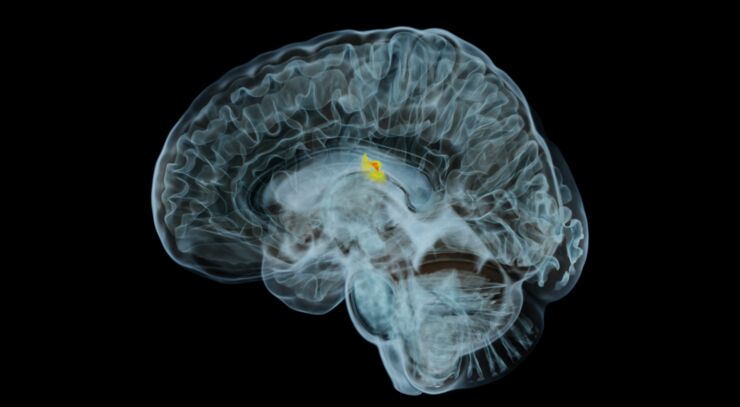
MR scan of tumor in a human brain Location

The workshop will take place at the auditorium K3 in the building Kåkenhus, Bredgatan 33, Campus Norrköping.

Registration

Free registration until 7 Dec 2022. Please fill in this form to register.

Contact persons

Lukáš Malý, lukas.maly@liu.se

Olof Svensson, olof.svensson@liu.se

Sponsors

LiU Cancer
Department of Science and Technology (LiU)

Organizers

George Baravdish, Vivianne Deniz, Rym Jaroudi, Tomas Johansson, Lukáš Malý, Olof Svensson

Programme

08:45-09:00 Welcome and opening
09:00-09:45 Peter Larsson (Region Östergötland)
09:45-10:30 Elin Nyman (LiU)
10:30-10:45 Coffee break
10:45-11:30 Rym Jaroudi (LiU)
11:30-12:15 Anders Eklund (LiU)
12:15-14:00 Lunch
14:00-14:45 Iuliana Toma-Dasu (SU & KI)
14:45-15:30 Fredrik Löfman (RaySearch Laboratories)
15:30-15:45 Coffee break
15:45-16:30 Mehdi Astararki (KTH)
16:30-17:15 Ivan Shabo (KI)
17:15-17:30 Closing

Abstracts

Mehdi Astararki - Machine Learning Methods for Oncological Image Analysis

Rapid advances in the field of medical imaging modalities resulted in capturing high-resolution anatomical, physiological, functional, and metabolic quantitative information from cancerous organs. The applications of medical imaging have become increasingly crucial in the clinical routines of oncology, providing screening, diagnosis, treatment monitoring, and non/minimally-invasive evaluation of disease prognosis. Considering the growing role of medical imaging data on one side and the challenges of manually examining such an abundance of data on the other side, the development of computerized tools to automatically or semi-automatically examine the image data has attracted considerable interest. Hence, a variety of machine learning tools have been developed for oncological image analysis, aiming to assist clinicians with repetitive tasks in their workflow. This presentation covers essential steps of oncological image analysis, including tumor detection/segmentation, followed by imaging biomarkers development for diagnosis, prognosis, and outcome predictions. 

Anders Eklund - Federated learning and synthetic images for brain tumor segmentation

Deep learning can today be used to automatically segment tu- mors and risk organs, which will save time in a clinical workflow. Training deep learning models require large datasets, which in medical imaging are difficult to obtain due to GDPR. In this presentation I will talk about how to create large datasets for training deep learning models, without sending sensitive information between hospitals. The first solution is federated le- arning, where several computers together train a segmentation network, by only sending updates of the model between each other. The second solution is to train a computer to generate realistic synthetic tumor images, which can be sent between hospitals as they don’t belong to a specific person (GD- PR does not apply).

Rym Jaroudi - Parameter identification and growth simulation in inverse mathematical models for brain tumours

A powerful tool in understanding cancer development is the use of mathematical models and simulations. In this talk, we discuss a well-established partial differential equation model of reaction-diffusion type for brain tumour growth. The model describes the change over time of the normalised tumour cell density at a given time and position of the brain region as a consequence of two biological phenomena: proliferation and diffusion. We study the inverse problems of locating the brain tumour source (origin) and identifying the response to treatment based on this model. Our approach consists in recovering the initial spatial distribution of the tumour cell density as well as the treatment parameter starting from a later state, which can be given by a medical image. Regularization methods are used to solve the inverse problems as a sequence of well-posed forward problems. This framework provides additional information about the initial location of the tumour and the treatment effect that can be used by neuro-oncologists in their decision-making process. An additional recent benefit of techniques developed in this research is the generation of synthetic data both forward and backward in time, which can be fed in as training data to improve AI based methods for tumour detection/classification.

Peter Larsson - An introduction to Radiotherapy; from History to Future, Challenges and Opportunities

The aim of this talk is to introduce the basics and to give a hint of the complexity of radiotherapy to scientists working in other fields and to indicate how mathematical modelling is used in radiotherapy today and where there are room for improvements. The goal for all curative radiothera- py is to use ionising radiation to deliver an absorbed dose to all cancer cells in the body, large enough to kill all of them without causing unacceptable harm to healthy tissue. In most cases, this is a very difficult task. The vast development in radiotherapy technology during the last 7 decades have led to increased survival and less side effects among the treated patients. The development in recent years rely on the advances in several different disci- plines. One of the most important being the use of mathematical modelling. The radiation source, the radiation transport and the human body have all been modelled leading to a more precise radiation treatment. However, de- spite all developments a lot of improvements are still needed. An example is how to handle movements during the radiotherapy session, especially for tumours located in the lungs or near the diaphragm. Another is modelling of the dose-response for different individuals.
The need for more research and development in radiotherapy in Sweden has recently been noticed on a national level and e.g. Cancerfonden has announced research founds specifically directed towards the field. Around 70 000 people get a cancer diagnose each year in Sweden, approximately half of them are treated with radiotherapy. The implementation of new techniques and better models in radiotherapy have the potential to cure or increase the quality of life for a large number of people.

Fredrik Löfman - Machine Learning in radiation therapy

Treatment plan generation in radiation therapy is a very data heavy and labor intensive process where machine learning can be applied to improve efficiency and consistency for certain workflow steps. We present two such machine learning applications that we have developed, medical image segmentation and treatment plan generation, and describe the choice of methods and present results. We discuss our experiences in developing and deploying machine learning applications for clinical use and the important topics of performance monitoring and life-cycle management of machine learning models.

Elin Nyman - Mechanistic modeling of cellular signaling networks relevant for cancer

Cancer cells can bypass intracellular control mechanisms for cell growth and cell death due to genetic alterations. The genetic alterations can be targeted with therapeutic agents, with some successful examples. However, resistance development is commonly observed over time. To understand and potentially prevent resistance development, intracellular signaling processes are key. I have developed mechanistic mathematical models, based on ordinary differential equations, for intracellular signaling processes in cancer cells. Important results from this work includes a framework to suggest new targets for therapeutic agents, including combinations of targets, that are predicted to circumvent resistance development for a specific genetic alteration.

Ivan Shabo - Tumor microenvironment, differentiation, and progression – from basic biology to clinical context

Carcinogenesis is a sophisticated biological process consisting of progressive changes in somatic cells from premalignant to malignant phenotype. Despite the vast accumulating knowledge in tumor biology, the origin of cancer, the mechanisms of invasive cancer cell selection, and the geno-phenotypes required for metastasis remain enigmatic. Moreover, it is now widely accepted that the malignant behavior of cancer does not depend solely on tumor cell biology. According to Paget's original “seed and soil” hypothesis, tumor morphology and invasive phenotypes are largely determined by selection in the host tissue microenvironment.
 
Cell differentiation is a process by which proliferating cells gradually acquire tissue-specific function and phenotype. During carcinogenesis, the cancer cells lose tissue-specific markers developing a de-differentiated state with increased proliferative capacity and plasticity. Moreover, de-differentiation has also been implicated in therapeutic resistance among several types of solid tumors.

Generally, a cancer diagnosis is based on i.a clinical signs, radiologic findings, immunohistopathology, and genetic analysis. The treatment and outcomes of cancer depend on several factors, like age and gender of the patient, tumor size, metastatic burden, as well as functional and immuno-genetic profile of the tumor. With increasing insight into cancer biology, the clinical assessment of cancer has become more complex, with various diagnostic modalities, patient stratification systems, and treatment options. Due to this challenging complexity, there is a need for new mathematical models and stratification tools that might facilitate the clinical assessment of individual patients or patient groups based on their predicted treatment response or risk of disease.
Iuliana Toma-Dasu - Modelling tumour oxygenation and its influence on the treatment outcome - from theory to applications in radiotherapy

The main problem in curing cancer resides in the different microenvironment in tumours than in normal tissues. All three components of tumour microenvironment, i.e. tumour vasculature, tumour oxygenation and tumour metabolism, contribute to the response of the tumour to a particular treatment. For radiation therapy in particular, tumour oxygenation is a very important factor that determines the biological effect of radiation. Tumour hypoxia, the lack of oxygen, is caused by a deficient vascular network and an increased interstitial pressure. Many studies have shown that poor tumour oxygenation is one of the main factors that determine the failure of radiation treatment. Therefore it is very important to account for the tumour hypoxia in the planning of the treatment in an attempt to identify the patients with poor oxygen supply to the tumours and to administer them tailored treatment strategies.

One alternative method to experimental work for characterizing quantitatively the tumour microenvironment is the theoretical simulation of the tissue based on measurable physical parameters. Thus, knowing the physical parameters of the tissue, one can use the equations that describe the fundamental physical processes in order to calculate theoretically the properties of the tissue microenvironment. In this respect, theoretical simulation of the tumour microenvironment is now the only available tool that may provide quantitative data for accurately describing tumour tissues. Thus, tissue oxygenation can be calculated starting from complex vascular arrangements and taking into consideration the oxygen diffusion into the tissue and its consumption at the cells. The results of the simulations could be used for modelling the tumour response to treatment or for investigating the efficiency of other measurements methods.

This presentation reviews important aspects that have been highlighted through the theoretical modelling of tissue oxygenation and in particular the relationship to vascular parameters. It also deals with the estimation of the efficiency of various measurement methods and the relationship between results obtained from different techniques. Particular attention has been given to resolution, averaging and other factors that may lead to systematic deviations of the measurement results. The role of the metabolic properties of the cells with decreased oxygenation was also taken into consideration for predictions of treatment outcome for full fractionated treatments. The findings stress the importance of incorporating hypoxia information into the biological modelling of tumour response for making clinical decisions. They also highlight the usefulness of theoretical simulation for evaluating the efficiency of strategies aimed to overcome the effects of tumour hypoxia.