“Sitting on a bus or in a meeting and not being able to break wind without embarrassing leakage. It’s not supposed to be like this, but some women have to cope with this after giving birth”, says consultant Sofia Pihl.
 Sofia Pihl, medical doctor. Photo: Ulrik SvedinShe describes in her thesis studies into how a new examination method has been implemented in perinatal care, and how obstetric personnel can assess ruptures more systematically.
“These assessments are difficult, and they must be made accurately all the time on an obstetric ward. So it’s important that effective procedures are available, together with simple technical aids”, she says.
Sophia brings out some ultrasound images and describes what happens.
“To describe it openly – this muscle helps us hold some sort of social control, so that you can withdraw from a group, for example, before breaking wind. The internal sphincter is kept under tension when it is inappropriate to release gas or faeces. And it helps us feel the difference between gas and faeces. So if this internal sphincter is ruptured, you lose social control. You cannot feel what is inside the bowels, and find it difficult to keep tight.”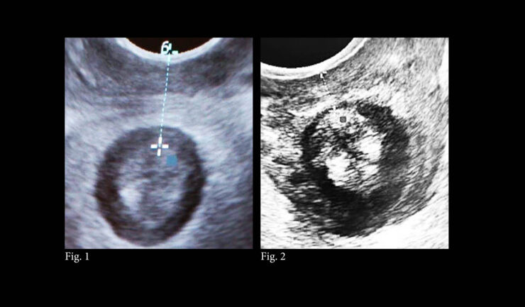 Ultrasound images of anal sphincter and perineal region. Midwives and other obstetric personnel can see the external sphincter without problems. But for the internal sphincter, they must learn how to search for ruptures using their fingers.
“It can, however, be difficult to reach a decision”, Sofia Pihl points out.
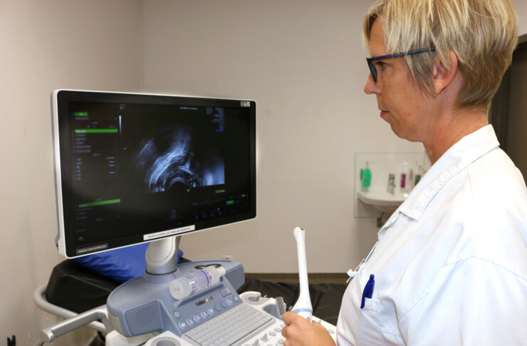
Personnel have used vaginal ultrasound probes for many years to examine the ovaries and uterus. They can also be used to examine the sphincter, using a method developed at Linköping University Hospital by Docent Eva Uustal. The ultrasound images show the rupture.
“If you point the probe backwards instead, you can see the muscles of the pelvic floor. Here you can see the sphincter, and determine whether it is circular or ruptured. And you can do it superficially, which means that the patient does not feel any pain.”
“It’s important to repair a rupture immediately. If it’s allowed to heal on its own, it is converted to connective tissue and becomes difficult to find.”
Sofia Pihl has investigated how long it takes for obstetric personnel to learn the new method, and has developed an equivalent scale to use when diagnosing the rupture.
“You have to do around five ultrasound examinations to learn the method. This is useful information, so we can know more about how the method can be introduced into the healthcare system. In the end, it’s actually a pretty simple method that uses existing equipment.”
Obstetric personnel already know a lot about ruptures. Traditionally the personnel mainly gain this through experience and through knowledge transfer from others.
“But we haven’t had an objective way of describing the ruptures. Instead, rather vague descriptions are used, such as: ‘A normal rupture. Sutured without difficulty.’ It can be difficult to obtain a good idea of what happened if it becomes necessary to examine the patient’s medical records later, if she experiences problems.”
The methods that have been introduced at Linköping University Hospital, under the direction of Eva Uustal, are based on both feeling with the fingers and using ultrasound, together with measurement of the perineum.
Sofia Pihl has compared the various methods and how ruptures are documented at different hospitals. An example:
“When an infant is delivered with its hand up by its cheek, for example, this is recorded in the notes, and we can also see that the mothers in these cases run a greater risk of damage to the internal sphincter. This knowledge can be used to remind the personnel that they should carry out a particularly thorough examination in these cases.”
In this work, Sofia Pihl has used the Swedish Perineal Laceration Registry (PLR), which was introduced in 2014. She has also examined how new methods can be introduced into the medical care system.
 Sofia Pihl, medical doctor. Photo: Ulrik Svedin“It requires stamina and clarity. The methods must be relevant, even if it’s 2.30 in the morning on a Saturday and the ward is fully occupied. I have described how it’s necessary to work at different levels and create structures that ensure that a new method really is used in practice. The equipment must be available, and the management must show willing. And there must be some committed people who get involved, and remind others about the new method.”
Sophia now wants to continue research into methods in perinatal care. But first, she is to go on a lecture tour.
“That’s right. I’m travelling to hospitals in Sweden to inform them about this method. We believe that it can make examination of all newly delivered mothers easier.”
(Translated by George Farrants)
“These assessments are difficult, and they must be made accurately all the time on an obstetric ward. So it’s important that effective procedures are available, together with simple technical aids”, she says.
Important functions
In 2019 in Sweden, approximately 3.2% of all first-time mothers experienced ruptures in the anal sphincter and perineal region after giving birth. Most of these women will not have any long-term problems. But the early discovery of such ruptures is important for the task of suturing the internal sphincter. This is a muscle with important everyday functions that many people do not even need to think about.Sophia brings out some ultrasound images and describes what happens.
“To describe it openly – this muscle helps us hold some sort of social control, so that you can withdraw from a group, for example, before breaking wind. The internal sphincter is kept under tension when it is inappropriate to release gas or faeces. And it helps us feel the difference between gas and faeces. So if this internal sphincter is ruptured, you lose social control. You cannot feel what is inside the bowels, and find it difficult to keep tight.”
Learning process
“It can, however, be difficult to reach a decision”, Sofia Pihl points out.
Personnel have used vaginal ultrasound probes for many years to examine the ovaries and uterus. They can also be used to examine the sphincter, using a method developed at Linköping University Hospital by Docent Eva Uustal. The ultrasound images show the rupture.
“If you point the probe backwards instead, you can see the muscles of the pelvic floor. Here you can see the sphincter, and determine whether it is circular or ruptured. And you can do it superficially, which means that the patient does not feel any pain.”
Immediate reparation
After giving birth, it can make a complete difference for a long time.“It’s important to repair a rupture immediately. If it’s allowed to heal on its own, it is converted to connective tissue and becomes difficult to find.”
Sofia Pihl has investigated how long it takes for obstetric personnel to learn the new method, and has developed an equivalent scale to use when diagnosing the rupture.
“You have to do around five ultrasound examinations to learn the method. This is useful information, so we can know more about how the method can be introduced into the healthcare system. In the end, it’s actually a pretty simple method that uses existing equipment.”
Obstetric personnel already know a lot about ruptures. Traditionally the personnel mainly gain this through experience and through knowledge transfer from others.
“But we haven’t had an objective way of describing the ruptures. Instead, rather vague descriptions are used, such as: ‘A normal rupture. Sutured without difficulty.’ It can be difficult to obtain a good idea of what happened if it becomes necessary to examine the patient’s medical records later, if she experiences problems.”
The methods that have been introduced at Linköping University Hospital, under the direction of Eva Uustal, are based on both feeling with the fingers and using ultrasound, together with measurement of the perineum.
How new methods are introduced
“This means that we have both an objective measurement and an ultrasound image. It gives much more reliable information, and a starting point for any subsequent care. When objective measures are taken, all personnel use the same language and descriptions of the ruptures.”Sofia Pihl has compared the various methods and how ruptures are documented at different hospitals. An example:
“When an infant is delivered with its hand up by its cheek, for example, this is recorded in the notes, and we can also see that the mothers in these cases run a greater risk of damage to the internal sphincter. This knowledge can be used to remind the personnel that they should carry out a particularly thorough examination in these cases.”
In this work, Sofia Pihl has used the Swedish Perineal Laceration Registry (PLR), which was introduced in 2014. She has also examined how new methods can be introduced into the medical care system.
Information to other hospitals
Sophia now wants to continue research into methods in perinatal care. But first, she is to go on a lecture tour.
“That’s right. I’m travelling to hospitals in Sweden to inform them about this method. We believe that it can make examination of all newly delivered mothers easier.”
(Translated by George Farrants)
Facts
Dissertation: Clinical and methodological aspects on perineal laceration diagnostics at childbirth
DOI: 10.3384/diss.diva-162580
Author: Sofia Pihl.
