Medical Image Science

Medical image science comprises techniques and processes intended to create images of a person for medical purposes. It reaches across disciplines such as radiology, endoscopy, microscopy, image processing and visualization.

Image from CMIV
Radiology means imaging the inside of the human body for the purpose of making a diagnosis. The term medical radiology includes diagnostic radiology as well as intervention - treatment guided by images.
 
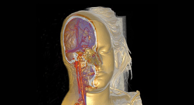
Development over the last few years of computed tomography, magnetic resonance imaging, and ultrasound and software for three-dimensional imaging offers entirely new opportunities for diagnosis and treatment. At the molecular and cellular level, the new techniques will make diagnosis possible before symptoms appear, and allow individually-adapted gene-based therapy with great precision. The surgery of the future will be bloodless and preserve healthy tissue.

Research

Two scientists are sitting infront of an MRI

Center for Medical Image Science and Visualization (CMIV)

CMIV conducts focused front-line research providing solutions to tomorrow’s clinical issues. The CMIV mission is to develop future methods and tools for image analysis and visualization for applications within health care and medical research.

Group of PhD students in the lecture hall

CMIV Research School

CMIV offers a doctoral program with both medical and technological entries and coherent research education. A basic principle for the program is a close connection between different disciplines i.e medicine, health, technology, and natural sciences.

Two researchers are talking

MRI-based Body Composition Analysis

Metabolic risk related to body-fat accumulation is strongly dependent on fat distribution. This project has developed a method for quantitative assessment of body composition that measures both fat distribution and muscle volume.

Research center

News

animated picture of a skeleton and photo of a skiing man.

Skiing becomes pure mechanics in his research

How do a skier's movements work? Which technique is the best? Biomechanics researcher Joakim Holmberg has studied this. Using mathematical calculations, he was able to follow how the force was distributed for each push of the ski poles.

Researchers discussing in front of a big screen displaying an image of a brain.

Advanced MRI technology detects changes in the brain after COVID-19

Researchers at LiU have examined the brains of 16 patients previously hospitalised for COVID-19 with persisting symptoms. Their findings can bring insights into the underlying mechanisms of persisting neurological problems after COVID-19.

male and female researchers discuss a CT scan.

Immune system ageing can be revealed by CT scan

Thymus may play a bigger role in the immune system of adults than was previously believed. With age, the glandular tissue in the thymus is replaced by fat, but the rate at which this happens is linked to sex, age and lifestyle factors.